How does the wound heal ?
1 BY growing 3D Cells with combination of herbs by increase intercellular signalling .
2 While Eradicating all antibiotic resistent Bacteria on Wounds without Anti biotics .
3 with Increase in WBC , T Cells, Fibroplasts , immunity .
4 Forming Collagen and intercellular granulating tissue capillaries Tissue muscle skin for closure and heal of diabetic foot ulcer .
1The acute response to injury involves the activation of the hemostatic cascade, which includes the release of growth factors and cytokines. 2 The second phase of wound healing involves the recruitment of neutrophils and establishes an inflammatory response. Macrophages recruited as part of the inflammatory response release additional growth factors and cytokines, which attract fibroblasts and endothelial cells to the site of injury.
3This third phase is marked by the proliferation of these cells and the deposition of extracellular matrix. 4 The final, or remodeling, phase of wound healing is characterized by keratinocyte migration into the healing wound site.
Applying . .my herbel medicine has carbon atoms equal to that of skin which cover the wound by absorption by skin . Enhancement of oxygen , exchange of gases ,circulation nutrition , metabolism ,growth division of cells at cellular level forming 3d cells thereby giving cell signaling to heal diabetic foot ulcer in high sugar diabetics , sterilizing all pathogenic bacteria to prevent amputation and death . grows nerve endings collagen muscle tissue without deformity as per genetic transcription in 6 weeks ,
Increase of Pituatary gland Growth hormone and Epidermal Growth factor , Fibroplast Growth factor , Platelet derived growth factor , high levels of exogenous peptides formed for cure in chronic wounds resulting from diabetes , for recirculation of blood to skin in the microscopic blood vessals in the skin so that neutraphils, macrophages regulates immune cell activation , fibroplast chemotaxis ,and vaso constriction of peripheral vascular system removal of bacterial toxins ,bacteria virulent factors , mmp cell debris this occurs by washing with hydrogen peroxide several times a day . Keratinocytes are formed from keratolinen(keratohyalic structures with profilaggrin ,dephosphorelation and proteolosis of profillagrin to fillagrin occurs within granular cells and keratinocytes move to form the horny layer upward i.e. Cornea Stratum . filaggrin functions as a glue to bind keratin filamints together . which break down to free amino acids to maintain skin hydration as lower new cells are formed .absence of keratin rises to skin dieseases and loss of moisture from cells .
Keratinocytes from corneocytes formed by proteins which mature from nuclei become large flat forming stratum corneum and in the upper layer with barrier lipids become rancid with fats under skin epidermis which protects skin against water loss stimulated by sun tanning and scavenger radicals , weak immunity .Throughout the 4 phases of wound healing, collagen performs the following functions in wound healing: • Guiding Function: Collagen fibers serve to guide fibroblasts. Fibroblasts migrate along a connective tissue matrix. • Chemotactic Properties: The large surface area available on collagen fibers can attract fibrogenic cells which help in healing. • Nucleation: Collagen, in the presence of certain neutral salt molecules can act as a nucleating agent causing formation of fibrillar structures.. • Hemostatic properties: Blood platelets interact with the collagen to make a hemostatic plug.
Top layer of the skin is the epidermis , keratinocyte activation, neutraphils terminates inflammation, regulates keratinocyte and endothelial cell growth ,inhibits proteases, activates leukocytes , stimulates nerve growth, keratinocyte proliferation, myofibroplast proliferation , to accelerate wound healing .
Healthy granular cells below epidermal layer produce involucrin loricin, keratolinene ,which are cross linked by trans glutaminases, to form gamma glutamyl lysine isopeptide bonds to form cell envelope .in their absence ulcers develop as skin becomes weak when inner epidermis is not growing for replacement above skin . hence we use agents as curcuminoides with soya lecithin for neutralizing the toxins produced , lysine , leucine , iso leucine, proline ,glycine , all consumed for avalibility with phosphotidyl choline, serine ( phospho lipids) to form new membrane structures below which develop to keratinocytes above .fatty acids as dry fruits softened in water is used as structurel proteins free fatty acids to wound closing with collagen growth . they make the layers tough so that there is no further deterioration and moisturizer used to keep the wound cured with sufficient moisture , .
Ceramides , fatty acids , cholesterol ,barrier lipids .make the stratum corneum . Intercellular granules , membrane bound lamellar granules, pro barrier lipids ,secreted interface of the horny and granular layer hydrolysed to form ceramides, free fatty acids in epidermis ,polysaccharides ex glycogen ,monosaccharide ex glucose store form of energy to meet cell division demand starts , folic acid by asparagus racemosa, beta carotene, betagluconone ,bee propalis , carotenoids astaxanthin ,tetra curcuminoides ,soya beans ,tulasi liquorice for anti inflammation are some neutraceuticals to be taken .
most of the body toxins as Endothelins ,Leucotrienes, chemokines ,Prostaglandins ,Protein kinases ,reactive oxygen species lead to inflammation and dissolve the skin and cells by forming elastase and collagenase enzymes which due to decrease in changes of numbers of neutrophils , wbc , dissolve the lysosomes , are washes out with hydrogen peroxide several times a day due to genetic repaire dna rna mitochondria .The transcription of genetic code is transmitted to other cells adjacent where the gentic transcription continues to more cells surrounding leading to life . hyaluriniridase chondroitin sulphate , arachidonic acid are consumed for nutrients and moisture for skin as vit e vit d etc food supplements for growth of new cells, substituting the dead cells, elasticity of skin increases and skin is grown . stimulation of immunity and wbc eradication of bacteria gram +and gram –ve leading to amputation prevented curing the wound by the bacteria which resides on the herb which is heat stable even at 400degc and consumes all residing pathogeneic bacteria . pino cytosis or phagocytosis starts hence infection cannot grow that reside on open wounds, so that sterile conditions are not needed ,formation of keratinocytes is enhanced ,as corneocytes grow below, Melanocytes ( pigmentation ),langherhan cells ( antigen),merkel cells ( sensory cells) appear with nerve micro capillaries , sweat glands, blood vessals , muscle ,connective tissue and all epidermis ,endodermis arise
Patients treated
the story of fisherwoman : the woman is 60 yrs old suffering from foot ulcer visited all famous hospitals hospital and I met her at diabetic physician dr .ch .padmavathi devi at padmavathi diabetic nursing home kothapet ,recognised by Novartis in Guntur for her services , .under her supervision the first trials were taken up . the patient is high diabetic and does not wear any slippers she is to roam daily without sandals . she visited number of doctors and first did not believe that I would give medicine as no doctor gave medicine and was afraid to use the medicine . she was given the required powder free for application 2-3times a day for external use the wound was washed several times with hydrogen peroxide and completely applied with powder .the necrosis tissue was cut by the doctor and treatment started .she used to eat fish epa dhea available ,
after 20 days the wound started healing to all amazement the wound closed inward she had the ulcer for more than two years treatment . there was no need of taking sterile measures after giving herbel medicines she did not wear chappals and she could go up the hill after treatment .
another photo of a poor washerman , the ulcer was 2cms depth and two cms width from three years ,he was afraid of amputation and there was no medicine . the ulcer healed off in 2 months with special herbel formula . watch the scar . there is no reoccurence of ulcers , pain ,plastic surgery, costly antibiotics ,and holds good from last 8 years .
this is due to growing 3d cells which have the best intercellular cell signaling bringing in life activity as originel due to genetic messaging as original .which is absent in plastic surgery hence reoccurrence of ulcer due to no equilibrium . herb consists of the number carbon atoms as skin hence absorbed easily .it also consists of important marriage or genetic combination of 2 herbs essential amino acid and fibrils to make collagen strong .the bacteria on the herbs destroys all bacteria on wounds . other Ingredients used top secret patent .
vitamin k2 with pentose sugar as ( Arabic gum pentose )or honey dew honey from Australia ,chitosan, caudate herb are used for prevention of bleeding wet wounds.
Our herb consists of fibrils for strengthening the wounds collagen fibre and hence .fibroplasts myofibroplasts , actin ,elastin, collagen bundles causing wound to contract and the wound is closed with epithelium and collagen granulating tissue, centrally.
Action of Hydrogen peroxide on washing wounds 5times-8times /day - Oxygen is vital for healing wounds. It is intricately involved in numerous biological processes including cell proliferation, angiogenesis, and protein synthesis, which are required for restoration of tissue function and integrity. Adequate wound tissue oxygenation can trigger healing responses and favorably influence the outcomes of other treatment modalities . A key moderator of normal wound healing is oxygen. Indeed, oxygen is a requirement for various processes in the healing of wounds including collagen deposition, epithiliazation, fibroplasia, angiogenesis, and resistance to infection. A limitation in the delivery of O2-rich blood to the wound tissue, therefore, impedes physiologic healing. The limitation of oxygen delivery to the wound is often multi-factorial; however, the end result is a hypoxic (oxygen deprived) wound microenvironment characterized by insufficient nutrient and oxygen delivery to the regenerating tissue. In this context, the state-of-wound tissue oxygenation is a major determinant of healing outcomes. tissue confronted with chronic, severe hypoxia does not survive. The same holds true for elevated wound tissue oxygen partial pressures where a moderate hyperoxic challenge can stimulate the production of growth factors and the formation of new blood vessels, whereas extreme hyperoxia can induce mitochondrial apoptosis, growth arrest, and oxidative stress via the formation of reactive oxygen species (ROS). Thus, the inherent complexity of the healing process mandates the precise combination of hypoxic cellular signaling and antioxidant defense mechanisms with adequate tissue oxygenation .
. The major role oxygen plays in many of the In the chronic wound microenvironment, there inevitably exists a substantial imbalance between the supply of oxygen and the high energy demand of the healing tissue. From a molecular standpoint, the key factors that propagate this imbalance include the following: (1) the increased utilization of oxygen by the hyper metabolic regenerating tissue, (2) the sustained and increased production of ROS by phagocytes (respiratory burst), and (3) reduction- oxidation (redox) signaling.1 Redox signaling and ROS generation is a process reliant on the NADPH oxidase (NOX) family of enzymes. The redox signals produced serve critical roles within thewound bed: (1) they provide the phagocytic immune cells with potent disinfecting capabilities, (2) in physiologic concentrations, they function as signaling molecules that mediate various responses, including cell proliferation, migration, differentiation, gene expression, and vascular tone.7,8 Although NOX-dependent O2 consumption is vital for appropriate redox signaling within the wound, it is important to understand that the optimal production of redox signals is imperative, as excessive ROS production can rapidly contribute toessential wound healing processes has also attracted considerable clinical interest, and investigations addressing the treatment of wound tissue hypoxia have yielded compelling data.4–6 In addition, scientific studies targeting the oxygen response signaling pathways within the wound tissue milieu are continuing to enhance our understanding of the biological and chemical pathways involved in the hypoxia sensing and response systems. This is of particular importance for the exploitation of new treatments that target hypoxia-response mechanisms within the healing tissue, thus making them useful in the wound clinic when combined with appropriate tissue oxygenation.
In the chronic wound microenvironment, there inevitably exists a substantial imbalance between the supply of oxygen and the high energy demand of the healing tissue. From a molecular standpoint, the key factors that propagate this imbalance include the following: (1) the increased utilization of oxygen by the hypermetabolic regenerating tissue, (2) the sustained and increased production of ROS by phagocytes (respiratory burst), and (3) reduction- oxidation (redox) signaling.1 Redox signaling and ROS generation is a process reliant on the NADPH oxidase (NOX) family of enzymes. The redox signals produced serve critical role within thewound bed: (1) they provide the phagocytic immune cellswith potent disinfecting capabilities, (2) in physiologic concentrations, they function as signaling molecules that mediate various responses, including cell proliferation, migration, differentiation, gene expression, and vascular tone.7,8 Although NOX-dependent O2 consumption is vital for appropriate redox signaling within the wound, it is important to understand that the optimal production of redox signals is imperative, as excessive ROS production can rapidly contribute to a hypoxic wound environment and cell death. Accordingly, the severely hypoxic wound environment will lack the essential O2 necessary for crucial redox signaling and appropriate wound healing.1 To mitigate the effects of the hypoxic environment, many cells use the hypoxia inducible factor (HIF) cascade, which activates multiple genes involved in cell proliferation, angiogenesis, and glucose metabolism. HIF-1 is a protein composed of two subunits:HIF-1a and HIF-1b. The latter is constitutively expressed within the cell, whereas the former contains the-a subunit tightly regulated by O2 dependent enzymes referred to as prolyl hydroxylase
domain enzymes (PHDs). Under normoxic (oxygen replete) conditions, the PHDs inactivate the HIF- 1a subunit. Conversely, hypoxic conditions inactivate the PHDs; stabilization of the HIF-1a subunit ensues, thus allowing for the formation of the HIF- 1a/HIF-1b protein complex, which then translocates into the nucleus. The HIF protein complex can then bind to hypoxia responsive elements within the promoter regions of various genes that are turned on in a hypoxic environment.
Wound healing is an angiogenesis dependent process reliant on blood vessels to deliver nutrients and oxygen to the regenerating tissue. The formation of new blood vessels (neovascularization) in the wound proceeds in two ways under the influence of angiogenic stimuli, such as vascular endothelial growth factor (VEGF)—the most potent factor for neovascularization: (1) angiogenesis, the process of new vessel sprouting from existing vessels and residing endothelial cells (ECs); (2) vasculogenesis, the recruitment and differentiation of circulating endothelial progenitor cells (EPCs) toform new vessels.5,10 Accordingly, the efficacy of oxygen therapy in the healing wound is, in part, mediated by the enhancement of the angiogenic response. Numerous in vivo studies have demonstrated increased VEGF expression in wounds treated with systemic oxygen therapy (HBOT), and in vitro studies have shown increased levels of VEGF mRNA in cultured cells involved in the healing process.5,11,12 Interestingly the increased VEGF expression in wounds treated with HBOT appears to be mediated, in part, by the stabilization of HIF, which subsequently promotes the transcription of the VEGF gene.10,13 It is important to note that there have been conflicting data with regard to HBOT, HIF, and wound healing. HBOT may affect HIF-1 expression depending on tissue type and the type of wound; that is, HIF-1 levels may differ in the acute healing phase compared with the late healing phase, or in ischemic wound healing.10,14 In addition to the localized induction of angiogenesis, HBOT has been shown to stimulate vasculogenesis by enhancing the production of bone marrow endothelial nitric oxide synthase (BMe- NOS); BMeNOS produces nitric oxide (NO). NO subsequently enhances the mobilization of EPCs, which home to the wound and contribute to the formation of neovessels.
Further, HBOT is involved in fibroblast activation, the up-regulation of growth factors, the reduction of the inflammatory response, and wound disinfection.6 It is these data that that lend support to the utilization of HBOT as an adjunctive care measure for diabetic lower extremity ulcers.16 HBOT remains a well-established, adjunctive treatment for diabetic lower extremity wounds refractory to standard care practices. The majority of the recent clinical outcome studies continue to provide strong and convincing evidence that adjunctive HBOT, in the setting of diabetes, decreases the risk of major amputation and markedly improves wound healing outcomes.6,16 However ,the effect of HBOT on chronic wounds associated with other pathologies (arterial ulcers, pressureulcers, and venous ulcers) requires further rigorous randomized trials to appropriately validate outcomes.6 Another promising therapy that enhances the oxygenation of cutaneous wounds is TOT. TOT is an O2 gas emulsion applied to the superficial wound tissue. Published data strongly suggest that TOT can generate a sustained increase in tissue oxygen levels; it can also enhance the angiogenic response in chronic human wounds by inducing VEGF production.5 Major advantages of TOT appear to be its independence of the wound microcirculation, its lower cost compared with systemic oxygen therapy, the lower risk of oxygen toxicity, and its relative simplicity of application and portability.
HIF is an integral part of the cell hypoxia sensing and response mechanism that promotes cell survival via the induction of numerous genes. In the context of wound healing, HIF-1 transcriptional target products have been shown to enhance angiogenesis by regulating EC survival, migration and proliferation, vascular smooth muscle cell migration and proliferation, and by mobilizing circulating endothelial progenitor/ stem cells to the periphery. HIF-1 has also been suggested to positively influence re-epithiliazation in the healing wound by increasing keratinocyte migration.
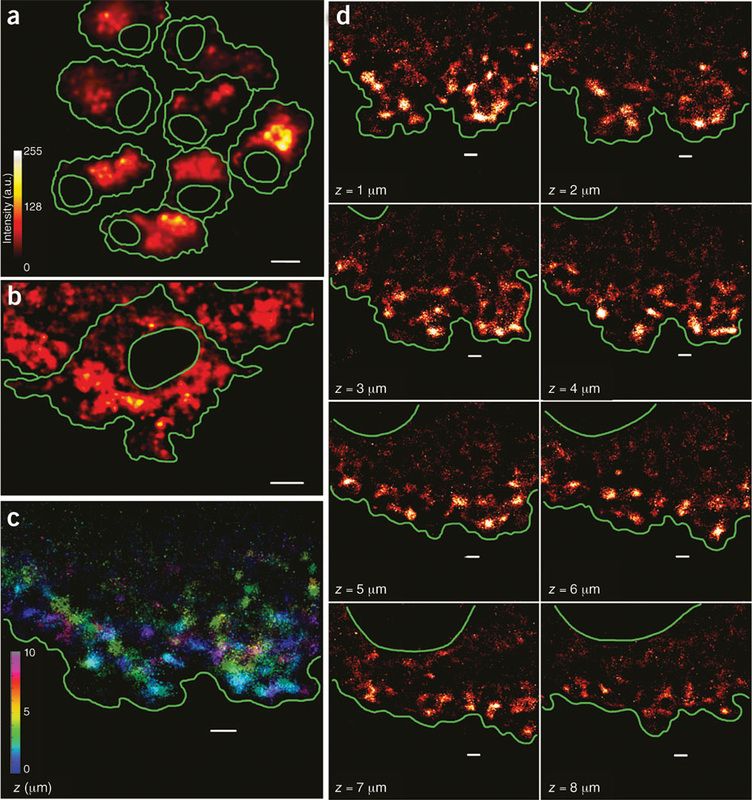
Searching for three-dimensionality biomarkers
Cytokines are soluble, low molecular weight, extracellular protein mediators that usually act at short range between neighboring cells. They are crucial intercellular regulators and mobilizers of cells engaged in innate and adaptive inflammatory host defenses, cell growth, differentiation, cell death, angiogenesis, and development and repair processes. They have been assigned to various family groups based on the structural homologies of their receptors and can be broadly classified into families such as colony stimulating factors, interleukins, interferons, transforming growth factors (TGF), tumor necrosis factors (TNF), platelet-derived growth factors (PDGF) and chemokines. Although cytokines have been extensively studied in the field of immunology and oncology, they have been overlooked by tissue engineers. However, these same small proteins might have the power to revolutionize the field. Although the evidence for their existence in 3D cultures is compelling,they have not yet been looked at as candidates for potential biomarkers. However, we suggest that they are the ideal family to explore for identification and validation follow-up studies for the following three main reasons.
First, when cells are transitioned from a 2D monolayer to a 3D microenvironment, they become surrounded by homotypic neighbors, forming a loosely bound disorganized aggregate. In vivo, such a scenario is encountered only during avascular tumorigenesis or early stages of inflammatory wound healing, which are both similar in nature and regulated by cytokines [29]. Therefore, in vitro, the cells growing in 3D related to any of those two models, depending on their type (malignant or primary, respectively), therefore explaining the upregulation of their cytokine levels.
Second, several 2D–3D comparative transcriptomic studies with cells from the four main tissue types (nerve, muscle, connective and epithelial) cultured on a variety of platforms, have reported the upregulation of cytokines and their receptors in 3D cultures. For example, Klapperich and Bertozzi [30] reported upregulation of seven cytokines [IL-8, chemokine (C-X-C motif) ligand (CXCL)-1, CXCL2, CXCL3, CXCL5, vascular endothelial growth factor (VEGF) and leukemia inhibitory facto (LIF)] by a human fetal lung fibroblast (IMR-90) cultured in a collagen– glycosaminoglycan (collagen/GAG) 3D mesh.
