DIABETIC FOOT ULCER

Background of
Diabetic foot ulcer
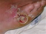
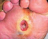
About 80 percent of foot ulcers occur on the bottom of insensate feet, or feet without feeling. They appear as shallow holes or craters which can vary in color, size and depth; they don't heal and may be extremely painful and in some cases give off an unpleasant smell. The medical term is gangrene. Expensive and time-consuming debridements, medications, antibiotics, therapy and skin grafts cannot correct the underlying process..As many as 50 percent of people with diabetes have a narrowing of the arteries supplying blood to the legs - a condition called peripheral vascular disease (PVD), a gradual buildup of fatty deposits on the walls of the circulatory system. As deposits, called atherosclerotic plaque, begin to clog the small vessels near the periphery of the system, tissues in the legs and feet can become starved for blood. Deprived of nutrients, oxygen, and germ-fighting blood cells, they become vulnerable to infection and ulceration. If the blood vessels to a portion of the legs or, more commonly, part of the feet become totally blocked, then the part of the body supplied by those blood vessels dies and gangrene occurs.
Ulcers are most often caused by repetitive mechanical stress that is not recognized by the patient because of peripheral neuropathy and the loss of sensation. A motor component to the peripheral neuropathy may lead to muscle atrophy, flexion deformity and/or abnormal gait, which, in turn, creates increased pressure points. Autonomic neuropathy causes dyshidrosis and dry skin, which is more prone to cracking. Finally, autonomic neuropathy may also be associated with arteriovenous shunting and altered skin perfusion .
The diabetic foot ulcer is a chronic wound and does not exhibit the orderly cascade of events which characterizes normal wound healing. Instead, diabetic ulcers appear to be “stuck” in the proliferative phase and are characterized by ongoing inflammation.
good glycemic control ,education on foot care ,appropriate costly foot wear , control of infection and early surgical intervention is needed in order to reduce the morbidity and mortality associated to dfu. Due to poly microbial infection and antibiotic resistence ,surgical intervention is considered .
So called diabetic foot is one specific example of gangrene that can be seen in long-standing complicated diabetes . Common in diabetics foot gangrene is typically caused by a combination of limb ischemia as the result of arterial occlusive disease, most commonly atherosclerosis , injury and poor healing, usually combined with a superimposed infection. Currently, the best of all possible standard medical diabetic foot treatments is revascularization (restoration) of the affected organ, which can reverse some of the effects of necrosis (tissue loss) and allow healing of the tissues Treatment other than revascularization, depending on the extent of tissue loss and location - digits, foot or hand - runs the gamut from allowing digits - toes or fingers - to auto-amputate (fall off) debridement (the surgical removal of necrotic, devitalized tissues) local care, usually with topical antibiotics, to amputation of foot or leg, depending on someone’s condition.
By the time the individual develops foot ulcers, his or her dire calls for medical help may be too late as most wounds are painless and not sensitive .
High levels of Glycosinated Haemoglobin are present in Diabetic Patients when excess of calories is taken with unknown high calorie foods as satuarated fats . The cells usually take minimum fixed known amount of Glucose for metabolism energy , which may vary in different cells , apart from storing glucose as glycogen in cells for time of need , fats formed from free fatty acids stored to form lipids which are transported in blood to cells and tissue for forming new cells .when there is no circulation fats forms are stored in the walls of arteries and micro capillaries . when there is increase in excess of glucose entry to cells more than that required by cells ,cells lock up themselves with inhibition of the operator on the gene . as they lock themselves , metabolism comes to an end , and circulation flow diminished, or pinocytosis and phagocytosis for exchange of nutrients metabolites stopped , oxygen to the cell closed ,leading to death of cells and genetic transcription happens to adjacent cells leading to open wounds of 2cm to 4cm, or gangrene of several cms on foot .
Disturbingly, research shows that diabetics who have had one lower limb amputated have a 50 percent mortality rate in the five years following the amputation. As well, they have 50 percent risk of developing a serious lesion in the second limb within two years, often leaving them immobile and putting them at risk of further complications from their diabetes. Remember, you're not invincible. Foot ulcer followed by amputation can get you. It can get anybody who has diabetes , dfu constitutes major source of morbidity and mortality among patients with diabetes mellitis
To this end, concentrated scientific efforts have continued to focus on the biological mechanisms that underlie wound complications with the ultimate goal of finding the most effective therapeutic modalities for afflicted patients
The risk of having an unattended wound really is infection. Wounds that are open for more than 30 days are much more likely to become infected, and once you develop an infection your risk of amputation goes much higher. To mitigate those risks, we actually will go in there and surgically remove the dead tissue. That’s called debridement. We’ll actually put wound gels on there to try and promote healing; and finally, we want to remove pressure on these wounds, which we call offloading. That can be done using crutches, a wheelchair, a walking boot, a cane, or even specialized shoes
most body toxins formed lead to inflammation and dissolve the skin and cells with hyalurinidase , elastase and collagenase enzymes which deteriorate elastin, actin, myosin hyaluronic acid, collagen , which due to decrease in changes of numbers of neutrophils , wbc , dissolve the lysosomes , and empty the hydrolases present in lysosomes into cells which digest cell membrane and cells , changes in immunity function and fibroplast chemotaxis , vaso constriction of peripheral vascular system results , increasing the ulcer.
Fibrillar Proteins, Structurel Proteins in form of long thin filaments , intracellular which are polymers of many basic protein molecules , provide contractile mechanisms of all muscles and to form microtubules, as nerve axons, mitotic spindles in growing cell division , extracellularly they are found in elastin fibres of collagen and connective tissues, blood vessals , ligaments, they are ,important for cell division , and growth which are absent in dfu .
arachidonic acid is dissolved to amino acids and water to give moisture to feet and replaced by cell growth below. chondriotin sulphate hyalorinidin which are inter active molecules decompose loosing water dryup the metabolism of cells come to end as moisture is needed for cell growth . dissolving arachidonic acid .
Globular proteins are soluble in cytoplasm and catalyse reactions with other substances in cell ex Glucose splitting to component , and combining with oxygen to form co2 , and water to give energy (atp). they are important in cell metabolism and absent in diabetic foot ulcer as there is no metabolism as glucose exceeds to spoil the cells . .Healthy granular cells below epidermal layer produce involucrin ,loricin , keratolinene ,which are cross linked by trans glutaminases , to form gamma glutamyl lysine isopeptide bonds to form cell envelope . in dfu all above are destroyed .
Lipids =fatty acids , to form structurel components of membranes are absent . glycerol storage form of energy to meet long term demands is stopped . free fatty acids absent .
Ceramides , fatty acids , cholesterol , barrier lipids .make the stratum corneum . Intercellular granules , membrane bound lamellar granules, pro barrier lipids ,secreted interface of the horny and granular layer hydrolysed to form Ceramides, free fatty acids, Polysaccharides ex glycogen , monosaccharide ex glucose is transferred to storage form of energy to meet short term demands in epidermis, all thease are used up by the cell at time of end of metabolism in cell , as already excess of glucose is present in blood and cell stops functioning due to closing of operon and new cells do not grow to replace the old cells damaged .
Cell protoplasm consists of water electrolytes , proteins, lipids, carbohydrates, water is present in cells except fat cells .70%-85% are ions as k.mg, so4, bicarbonate, na, ca, cl, which is linked to chemical reactions and maintenence of substances dissolved intracellular and extracellular in normal skin . thease are sent to the growing cells by increasing circulation to the effected part by increasing, wbs, the mechanism of transport does not work due to closed operon . When tissue is damaged histamine is derived from mast cells and basophils under inflammation causing vasodilation , arterioles increase capillary porosity allowing leakage of plasma fluid protein into tissue causing edema , k, mag, acetate citrate, increase of co2 , leading to vasodilation .
Pinocytosis when cell membrane becomes weak and breaks up 3% of The damaged portion of the membrane forms a pit and it invaginates inward and fills with necessary fibrillar protein surrounding the invaginated pit causes its border to close over attached protein with extracellular fluid forming pinocytic vesicle 100-200 nm with proteins transported to inside cytoplasm of cell which requires energy from atp .to attach to protein receptor on skin called pits , beneath is a lattice work of fibrillar proteins called clathrin and proteins, including contractile filaments of actin and myosin for contracting and forming pit which are absentin dfu .
Phagocytosis by tissue macrophages , wbc, when a bacteria dead cell or tissue debris binds with receptor on the surface of phagocyte the edges of the membrane invaginate and surrounds particle to close as phagocyte which are transported out of cell . Macrophages form pinocytic vescicles of 200 nm diameter to send intercellular metabolites ,and radicals to transport exocellularly by phagocytosis controlled as per Genetic original code transcription . Larger vesicles pinch off from the endoplasmic reticulam and fuse with golgi to form lysosomes secretory vesicles .which is absent in dfu as the metabolism stops .presence of calcium ion in the extracellular fluid probably react with protein filaments beneath pits to provide force to pinch the vesicles from cell membrane into cytosol .this is the basic feeding of proteins to form aminoacids , polysaccharides ,strength to skin ,to neutralize free radicals formed .. as collagen is formed it is deteriorated on surface of skin to form amino acid and water useful for moisturizing skin. Most of collagen is destroyed by Hyalurinidase ,Collagenase , Elastase enzyme actions on collagen , arachidonic acid , hyaluronic acid . . new collagen is formed from arachidonic acid below to replace the collagen destroyed .which isabsent in dfu .
When cell is damaged in dfu lysosomes ( used for digestion of proteins and debris for transport out of cells ) inside cell , rupture to give off hydrolase into cells to digest the cell contents itself if the damage is severe with toxins and radicals , the hydrolase released begin digestion of protein carbohydrate lipids and other substances inside cells , the product is aminoacids , glucose phosphatase that can diffuse through the membrane of the vesicle to the cytoplasm ordinarily. what is left of body represents undigested vesicle ,this is mostly excreted through the cell membrane by exocytosis , by metabolism which is absent in dfu .
Many other carbohydrates compounds called proteoglycans (mainly carbohydrate substances bound to small protein cores,) loosly attached to outer surface of cells forming loose carbohydrates Coat called Glycol calyx . the glycol calyx of some cells attaches to the glycol calyx of other cells ,and many other carbohydrates act as receptor substances for binding hormones as insulin absent in dfu .
Five special ingredients Endoplasmic reticulum, Golgi apparatus ,Mitochondria, Lysosomes, Peroxisomes, Ribosomes , are found on the surface of Granular Endoplasmic Reticulum which are composed of proteins and important in synthesis of new protein molecules . The agranular endoplasmic reticulum responsible for synthesis of lipid and enzymatic process inside cell are responsible for cell synthesis growth and during Absence of endoplasmic reticulum , golgi bodies , phospholipids , cholesterol , synthesis of essential enzymes , absence of glycogen breakdown for energy and absence of essential enzymes for detoxification of drugs that damage cells by process of coagulation , hydrolysis exists dfu results ..
Glycocalyx membrane carbohydrates ,occur with proteins or lipids in form of glycolipids, and glycol proteins .1/10 th of the membrane lipid molecules are glycolipids ,the glycoportion of thease molecules almost invariable protrude outside cell dangling outward from the cell surface .all thease are absent in dfu .
Conjugation with hyaluronic acid, large polysaccharides polymers bound with small amount of proteins as hyaluronic aicd , chondroitin sulphate , which form components of proteoglycan secreted in mucosa or other glandular secretions to form major, components of the ground substance in interstitial spaces acting as filler between collagen and fibres and cells Which is absent in cells with a closed operon damaged rna golgi ,resulting no metabolism and respiration ,exocytosis , pinocytosis resulting in ulcers . also present are Lipids that are soluble in fat soluble solvents as (phospholipids of soya lecithin,) cholesterol to form 2% of cell , some cells contain 95% of cell mass as tri glycerides .
Peroxisomes formed by self replication combining oxygen to hydroxide ions from intracellular chemicals to form hydrogen peroxide which is highly oxidizing and used with catalase to oxidise poisonous substances . cells without mitochondria are unabled to extract significiant energy from nutrients to cell which delays pinocytosis ,phagocytosis and cell death .
oxygen combines with all break down products of carbohydrates fat protein ,to release energy for cell function and all cells deliver end products to extracellular fluids ,almost all cells also have the ability to grow and generate new cells until supply is replenished .there is intracellular fluid inside cells ,as blood passes through capillaries continuous exchange of extracellular fluid occurs between plasm portion of the blood and the interstitial fluid that fills the spaces between cells .the capillaries are permeable to most molecules in plasma of blood which can diffuse back between blood and tissue spaces .wounds are deprived of oxygen .
.all cells are located within radious of 50 micrometres from capillary due to kinetic motion of molecules the extra cellular fluid everywhere in the body both of plasma and interstitial spaces maintaining complete homogeneity of fluids everywhere in the body .this is absent in dfu due to capillaries constricted no blood flow. Proteins and Polypeptide Hormones synthesized in the endoplasmic reticulum of different endocrine cells as pre hormones are inactive , cleared in endoplasmic reticulum, transferred to golgi for packaging into secretory vesicles, the vesicles are stored within cytoplasm and many are bound to cell membrane until their secretion is neccessary .
Decrease in Epidermal Growth factor , Fibroplast Growth factor , Platelet derived Growth factor , low levels of Exogenous Peptides, Autonomic Neuropathy, Pseudomotor Dysfunction ,decrease of vascular flow , within the skin of the sole of the foot leading to dry foot skin . there is no pino cytosis or phagocytosis hence infection can grow from bacteria g-ve and gram + ve , that reside on open wounds, increasing more so that sterile conditions have to be maintained with excess dressing skin , In diabetic foot ulcer formation keratinocytes is diminished ,as there are no corneocytes below, Melanocytes ( pigmentation ),langherhan cells ( antigen),merkel cells ( sensory cells) are lost .
INNOVATION
The closure of a full-thickness wound contraction is a slow process and not commonly used. Skin grafting is fast and effective in closing a defect. However, grafting, a surgical procedure, creates a new wound that causes discomfort as plastic surgery will lead to 2d cells and poses cosmetic issues. New therapies that lead to faster wound contraction should reduce the need for grafting. The development of more efficient wound contraction, possibly by enhancing the rate of collagen organization that would hasten the rate of contraction and eliminate both a surgical procedure and a secondary defect and scar, would be welcome.
Answering these questions requires the knowledge and expertise of many disciplines: most notably, cell biology and bioengineering. Health care providers within the clinic represent multiple disciplines, including vascular surgery, plastic surgery and reconstructive surgery, dermatology, orthopaedic foot and ankle surgery, hyper- baric medicine, and wound/ostomy nursing podiatry . Combining cells with scaffolding materials to generate functional tissue constructs describes tissue engineering at its most basic level.
TABLE 6 bacteria resitent costly anti biotics used daily to prevent infection not cureSelected Empiric Antibiotic Regimens for Diabetic Foot Ulcers
Scenario
Drug of choice
Alternatives*
Mild to moderate, localized cellulitis (outpatient)
Dicloxacillin (Pathocil)
Cephalexin (Keflex); amoxicillin/clavulanate potassium (Augmentin); oral clindamycin (Cleocin)
Moderate to severe cellulitis (inpatient)
Nafcillin (Unipen) or oxacillin
Cefazolin (Ancef); ampicillin/sulbactam (Unasyn); clindamycin IV; vancomycin (Vancocin)
Moderate to severe cellulitis with ischemia or significant local necrosis
Ampicillin/sulbactam
Ticarcillin/clavulanate (Timentin); piperacillin/tazobactam (Zosyn); clindamycin plus ciprofloxacin (Cipro); ceftazidime (Fortaz) or cefepime (Maxipime) or cefotaxime (Claforan) or ceftriaxone (Rocephin) plus metronidazole (Flagyl); cefazolin (for Staphylococcus aureus); nafcillin (Unipen); oxacillin
Life- or limb-threatening infection
Ticarcillin/clavulanate or piperacillin/tazobactam, with or without an aminoglycoside
Clindamycin plus ciprofloxacin or tobramycin (Nebcin); clindamycin plus ceftazidime or cefepime or cefotaxime or ceftriaxone; imipenem/cilastin (Primaxin) or meropenem (Merrem); vancomycin plus aztreonam (Azactam) plus metronidazole; vancomycin plus cefepime; ceftazidime plus metronidazole
IV = intravenous.
Besides continuing treatment for his diabetes, the person with a beginning ulceration to the foot has a war to wage against bacteria that will invade the compromised area. Staph aureus and strep, normal skin flora, most likely will cause the problem .Diabetic Foot Wounds
Causes of Venous and Arterial On the leg, venous and arterial ulcers are often caused by:
- Poor circulation
- Venous insufficiency
- Blood clots
- Hypertension
- Diabetes
- Kidney failure
- Inflammatory diseases
- Medications
- Infections
- Diagnosis
- X-rays
- MRIs
- CT scans
- Other non-invasive tests
- Compression
- Wound debridement (removal of tissue)
- Advanced wound dressing
- Surgery
- Bioengineered skin substitutes
- Vacuum-assisted closure